When deaths are violent, suspicious, sudden, and unexpected, a Medical Examiner performs a post-mortem examination. Medical examiners specialize in forensic knowledge and rely on this during their work. In addition to studying cadavers, they are also trained in toxicology, DNA technology and forensic serology (blood analysis). Pulling from each area of knowledge, a medical examiner is expert in determining a cause of death. This information can help law enforcement crack a case and is crucial to their ability to track criminals in the event of a homicide or other related events.
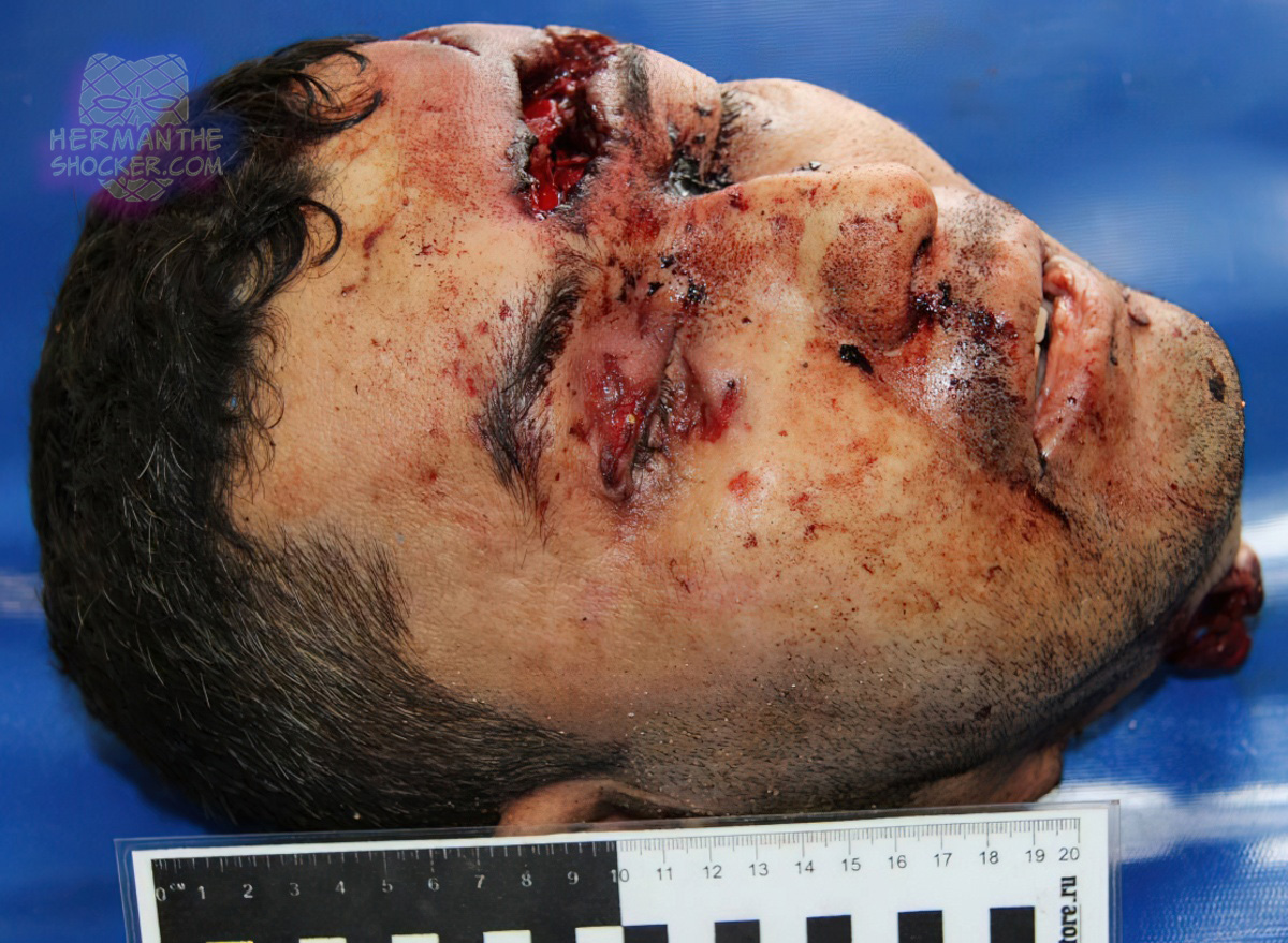
This happened in Russia. Nine parts of a dismembered human corpse were submitted for forensic examination: a head, right forearm, right hand, left forearm, left hand, right shin, right foot, left shin, left foot. All the parts had brownish-gray sandy soil on them. The parts were unclothed and without shoes. The head had been cut at the neck. There are lacerations on the forehead from a blunt force trauma.
The right forearm is separated from above at the level of the elbow joint, the plane of separation is directed obliquely horizontally, from behind to front and somewhat from top to bottom. The edges of the cut-off on the skin along the posterior surface of the joint are relatively even. No foreign particles were found on the soft tissues and skin in the area of dissection. From below, the forearm is separated at the level of the wrist joint, the plane of separation is horizontal. The cut-off edges on the skin along the palmar surface are relatively even, not furrowed; on the dorsal surface, they are finely serrated, with the formation of slightly furrowed sharp-angled teeth up to 0.5 cm high, partially exfoliated from the underlying tissue. The skin of the forearm is white, with black hair on the back surface, without additional damage, clean, without scars, tattoos, birthmarks, cadaveric spots on the skin are not distinguishable. There are no signs of decay, drying, or animal scavenging.
The right hand is separated at the level of the wrist joint, when compared with the right forearm, the planes of separation of the soft tissues and bones of the forearm completely coincide, without defects. Soft tissues in the plane of separation were slightly dried up, without hemorrhages. No foreign particles were found on the soft tissues and skin in the area of dissection. The skin of the hand is white, with black hair on the back surface, without additional damage, clean, without scars, tattoos, birthmarks, cadaveric spots on the skin are not distinguishable. There are no signs of decay, drying, or animal scavenging. There are no signs of maceration on the skin of the hand. There are no blood deposits on the palmar surface of the hand.
The left forearm is separated from above at the level of the elbow joint, the plane of separation is directed obliquely horizontally, from behind to front and somewhat from top to bottom. The cutoff edges on the skin along the posterior and outer surface of the joint are relatively even, not scaly, on the anterointernal surface they are finely serrated, not scaly, with the formation of acute-angled teeth up to 0.5 cm high, partially exfoliated from the underlying tissue. The subcutaneous tissue, muscles and tendons are cut at the same level in the plane of dissection. Soft tissues in the plane of separation were slightly dried up, without hemorrhages. Foreign particles on the soft tissues and on the skin in the area of dissection were not found.
The left hand is separated at the level of the wrist joint, when compared with the left forearm, the planes of separation of the soft tissues and bones of the forearm completely coincide, without defects. The soft tissues in the plane of separation were dried up, without hemorrhages. Foreign particles on the soft tissues and on the skin in the area of dissection were not found. The skin of the hand is white, with black hair on the back surface, clean, without scars, tattoos, birthmarks, cadaveric spots on the skin are not distinguishable. There are no signs of decay, drying, or animal scavenging. On the dorsal surface of the hand, in the projection of the bases of the 2nd-3rd metacarpal bones, there is a linear scratch 2.5 cm long directed at the numbers 1 and 7 of the dial with a yellowish-pink dried out sinking surface. On the palmar surface of the hand are dried overlays of blood in the form of adherent clots, small spots and single point splashes, without distinguishable streaks. A careful scraping of dried traces of blood from the palmar surface submitted to investigators.
The right shin is separated from above at the level of the knee joint, the plane of separation is directed horizontally in the anterior half of the joint, in the posterior half it is directed obliquely horizontally from front to back and from top to bottom. The edges of the detachment on the anterior, inner and lateral surface of the joint are relatively even, not shriveled. On the posterior surface, finely dentate, with the formation of small pointed teeth up to 0.8 cm high, not barred. The joint capsule is divided in front – at the level of the joint space, in the plane of separation – the articular surface of the tibial condyles, in the back of the joint – an obliquely horizontal cut of the posterior metaepiphyseal sections of the tibial condyles, on the cartilaginous part of which horizontal linear parallel grooves in the form of traces are visible. There is no blood in the cavity of the divided joint. Subcutaneous tissue and muscles are cut at the same level in the plane of dissection. From below, the lower leg is separated at the level of the ankle joint, horizontally. The cut-off edge on the skin is relatively even, not furrowed. Subcutaneous tissue, muscles and tendons are transversely crossed at the same level in the plane of dissection.
The right foot is separated transversely at the level of the ankle joint, 9 cm from the plantar surface, with sawing of the lower metaphyseal sections of the tibia and fibula, the cut passes through the cancellous bone. When compared with the right lower leg, the planes of separation of the soft tissues and bones of the lower leg completely coincide, without defects. The soft tissues in the plane of separation were dried up, without hemorrhages. Foreign particles on the soft tissues and on the skin in the area of dissection were not found. The nails are convex, of medium length, neatly trimmed. There are no blood deposits on the dorsal and plantar surfaces of the foot.
The left shin is separated from above at the level of the knee joint, the plane of separation is directed obliquely horizontally from front to back and from top to bottom. The edges of the detachment on the anterior surface of the joint are relatively even, not shriveled. On the outer, inner and posterior surface, finely serrated, with the formation of small pointed teeth up to 0.8 cm high, not barred. On the posterior surface, the edge of the detachment forms a large acute-angled flap upwards, 5 cm high, 3 cm at the base, with finely serrated margins slightly pitted. Subcutaneous tissue and muscles are cut at the same level in the plane of dissection. Soft tissues in the plane of separation were slightly dried up, without hemorrhages. From below, the lower leg is separated at the level of the ankle joint, horizontally. The cut-off edge on the skin is relatively even, not furrowed. Subcutaneous tissue, muscles and tendons are transversely crossed at the same level in the plane of dissection. On the anterior surface of the left lower leg at the knee joint, immediately below the edge of the dissection, there is an oval vertically oriented abrasion 1.5×1 cm with a brownish dense protruding surface exfoliating at the edges (old abrasion).
The left foot is separated transversely at the level of the ankle joint, 11 cm from the plantar surface, with sawing of the lower metaphyseal sections of the tibia and fibula, the cut passes through the cancellous bone. When compared with the left lower leg, the planes of separation of the soft tissues and bones of the lower leg completely coincide, without defects. The soft tissues in the plane of separation were dried up, without hemorrhages. Foreign particles on the soft tissues and on the skin in the area of dissection were not found. There are no blood deposits on the dorsal and plantar surfaces of the foot.
For internal examination a longitudinal incisions were made along the forearms, back surface of the hands, shins and back surface of the feet with separation of soft tissues. The brain was examined. Hemorrhages in the soft tissues of the extremities, including in the projection of a scratch on the dorsum of the left hand, abrasions of the left leg near the knee joint, were not found. To determine ethyl alcohol, skeletal muscles with markings were taken for forensic chemical examination. Samples of skeletal muscles were sent to the forensic biological laboratory to determine the group properties of blood.
Latest posts


