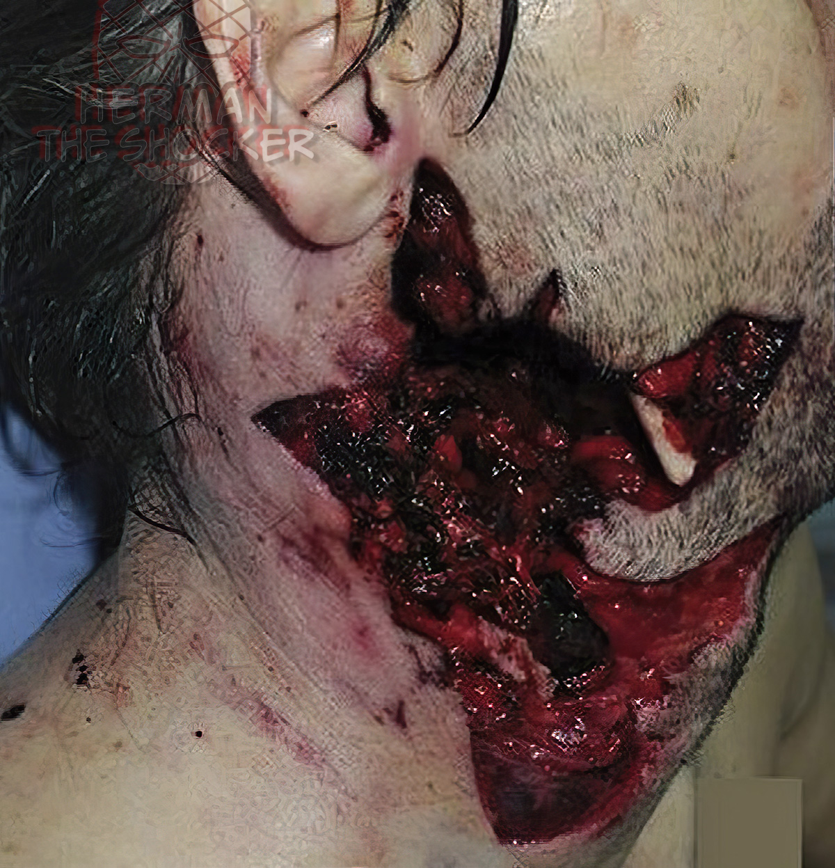
This middle-aged individual was involved in an altercation with another male relating to the manufacture of amphetamines. Circumstantial evidence indicated that the deceased was shot with a 12-gauge, bolt-action, sawn-off shotgun. The firearm was not located. The cause of death was from a gunshot wound to the neck.
On the left side of the neck, there was a gunshot entrance wound that was roughly circular measuring approximately 1.5 cm in diameter. This wound was associated with extensive laceration of the left external ear with disruption of the lobe and tragus. There were no satellite pellet marks on the adjacent skin. There was no muzzle imprint or stippling. Although there was the suggestion of irregularity of the wound margin, there was no well-defined scalloping. In its depth, the wound was associated with fracturing of the left mandible.
Situated on the right side of the neck extending from a point immediately anterior to the right earlobe was a gaping wound that measured 14cm superoinferiorly × 13cm anteroposteriorly (with the head in the anatomical position). This was a gunshot exit wound. The margin of the wound was irregular, comprising a number of discrete lacerations. There was no blackening of wound margin, muzzle imprint, or stippling. There was a laceration to the upper lip on the left approximately 2 cm from the midline, which is full thickness and probably associated with the force from the projectile and bony damage expanding the soft tissue around the projectile track.
The following fractures were identified at autopsy:
- Skull: there was a hinge fracture at the junction of the right middle and posterior cranial fossae extending from the right petrous temporal bone through the sella turcica.
- Spinal column: there was a comminuted fracture-dislocation of the atlantooccipital joint with comminuted fracturing of the atlas.
- Mandible: left and right bodies.
- Nasal bone.
- Larynx: the left and right superior cornua (horns) of the thyroid cartilage were fractured as were the left and right greater horns of the hyoid bone. The fracture of the left greater horn of the hyoid bone involved the tip, and there was minimal hemorrhage. The fracture of the right greater horn involved the junction of the proximal one-third and distal two-thirds and was related to the gunshot wound track.
Given the absence of muzzle imprint and satellite pellet marks, the range was estimated to be close but not contact. Precise determination of range would depend on test firing of the culprit firearm. Circumstantial evidence indicated that the deceased was shot with a 12-gauge, bolt-action, sawn-off shotgun. The firearm was not located.
Postmortem CT showed the extent of the fracturing to the inferior head and neck, particularly the right side, and the hinge fracture. A large number of gunshot pellets of approximate diameter 3mm were located within the wound track and adjacent to the cervical spine and mandibular fractures.
Latest posts


