Swelling of the scrotum and abdomen occurs with putrefaction as gases accumulate from bacterial action. Numerous maggots can also be seen on the surface of the body. Usually the first external naked-eye sign of putrefaction is discoloration of the lower abdominal wall, most often in the right iliac fossa where the bacteria-laden caecum lies fairly superficially. Direct spread of organisms from the bowel into the tissues of the abdominal wall breaks down haemoglobin into sulphaemoglobin and other pigmented substances. This discoloration spreads progressively over the abdomen, which in the later phase begins to become distended with gas. At about this time, more generalized spread of bacteria begins to discolour the more moist tissues.

Fig.1 Swelling of the scrotum and abdomen from putrefaction and accumulation of gases.
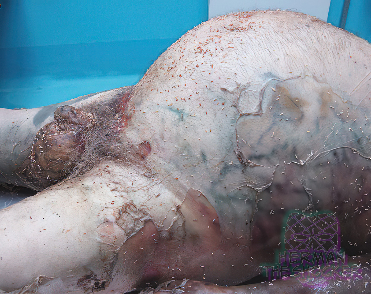
The putrefactive bacteria, which largely originate in the intestines and lungs, spread most easily in fluid so they tend to colonize the venous system, haemolysing the blood that stains the vessel walls and adjacent tissues. This gives rise to ‘marbling’, a branching outline of arborescent red, then greenish pattern in the skin, seen most clearly on the thighs, sides of the abdomen, and chest and shoulders. At or even before the stage of marbling, skin blisters may appear, at first on the lower surfaces of trunk and thighs where hypostatic oedema has loaded the tissues with fluid.
Gas formation will now become marked, with increased tension in the abdomen. The scrotum and penis may swell up to remarkable size and the neck and face will become grotesquely bloated, making visual identification difficult or impossible. The pressure may cause the eye globes and tongue to protrude. Purging of urine and faeces may occur due to the intra-abdominal pressure and, occasionally, a uterine prolapse may be extruded. There are recorded instances of pregnant women having a macabre post-mortem ‘delivery’ of the fetus, from the same cause.

Latest posts


