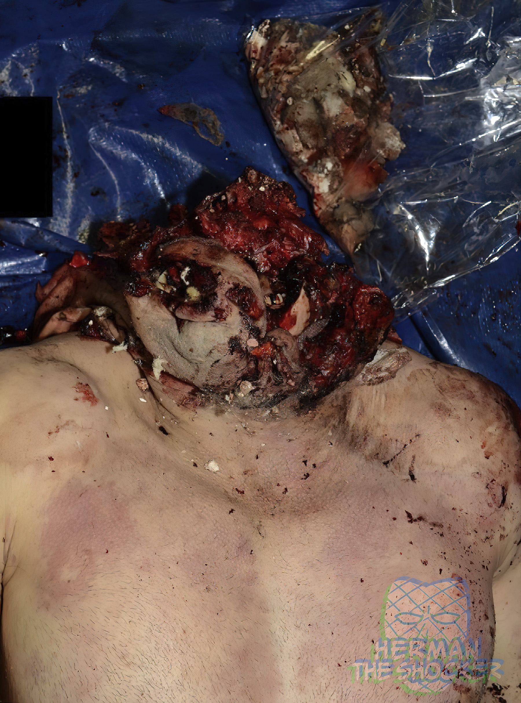
This male individual placed an explosive detonator in his mouth. There was extensive destruction of the bone and soft tissue of the cranium. The scalp and fragments of bone and cerebral tissue were received separately from the body in a bag. There was black discoloration of the skin at the margins of the complex lacerations and black discoloration of the dorsum of the tongue. There was a complex laceration at the neck that merged with the craniocervical injury complex.
The massive craniocerebral destruction is common in non-accidental deaths involving explosives. Explosives are, however, also commonly used in armed conflict. Blast injuries have been well documented. Taking into account where the explosion/blast occurs (e.g., confined space or open air), an examination of injury patterns with regard to the degree of fragmentation of the body may provide information about the distance of the victim/s from the point of explosion/blast.

Fig.1 Anterior view of the head and chest showing extensive trauma to the head.

Fig.2 Superior view of the head and chest showing extensive trauma to the head.
Postmortem CT showed extensive small fragmentation of the entire cranial vault and mandible. The majority of the cervical vertebrae were intact with the exception of the 6th cervical vertebra, which had a vertical fracture of the body. Although vertical split injuries may be seen in association with flexion teardrop fractures, equally there may be no associated abnormalities (as is the case here), and these have been described as compression injuries.

Fig.3 VR images of the skull showing extensive fracturing.

Fig.4 VR images of the skull showing extensive fracturing.

Fig.5 VR images of the skull showing extensive fracturing.

Fig.6 Coronal reconstruction of the neck and upper chest showing a vertical split fracture of the body of the 6th cervical vertebra (red arrow).

Latest posts


