I unfortunately don’t have the backstory here, other than this is from a case of postmortem animal interference, somewhere in Panama. I’ve added some general info instead.

Fig.1
Postmortem animal depredation is a substantial part of the taphonomic processes a body undergoes after death and is a well-known phenomenon to forensic death investigators, the morphological appearance of these injuries can be misinterpreted by police officers investigating the suspected crime scene or by the consulted medical practitioner.
A broad range of carnivores can be involved in the postmortem destruction of corpses located in open spaces or indoors (e.g. wild animals such as foxes and big cats or domestic animals such as dogs and cats). The wound margins caused by carnivores often appear more regular than those caused by rodents and V-shaped or rhomboid punctured wounds are often seen upon the intact skin in the immediate vicinity to the actual wound margins. Such stab wound-like defects represent canine tooth marks of carnivore origin. An additional criterion for animal depredation by carnivores is the presence of claw-induced linear scratch-type abrasions in the vicinity of the damaged skin areas.

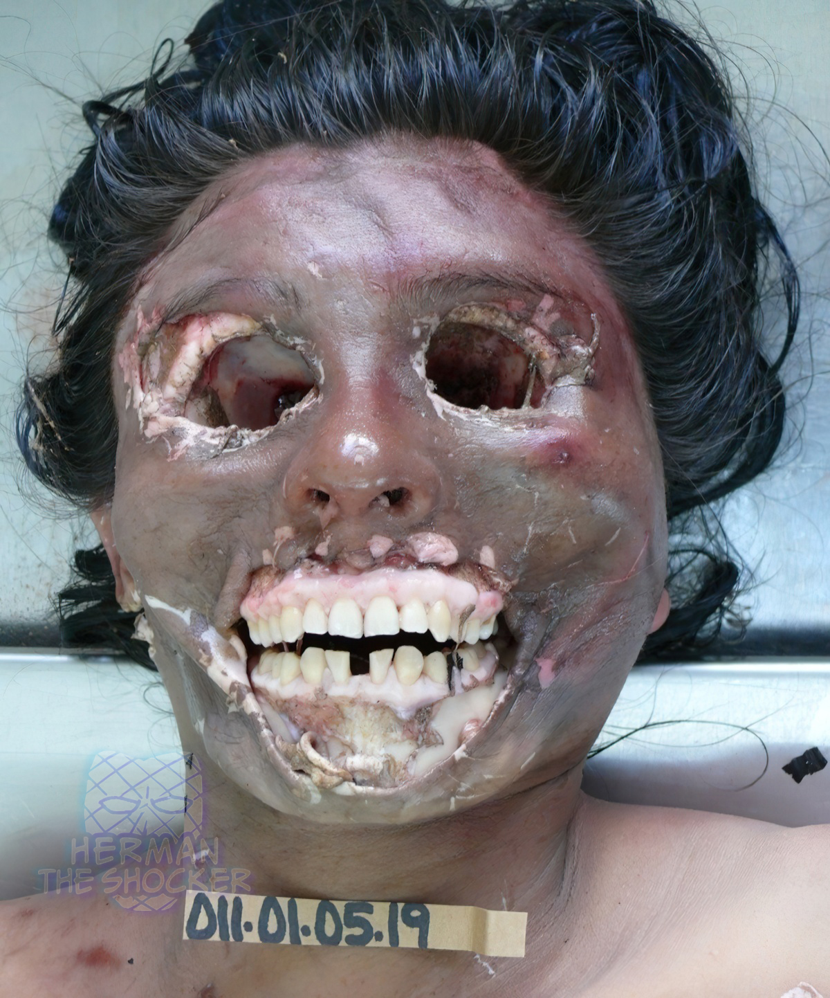
Fig.2
Determination of rodent species from the morphological appearance of damage to soft tissue and bones is often unreliable. In cases of postmortem rodent interference, the appearance of distinct parallel cutaneous lacerations in the margins of damaged skin can be attributed to the animals masticatory apparatus, namely the pair of incisors.
Frequently the face, hands and legs are destroyed by postmortem animal interference as they are unclothed and thus easily accessible. However there are cases where such lesions having been noted in other areas of the body which have been covered by the cloths including genitals but they are rare.

Fig.3
Death investigators should be aware of the following while undertaking a detailed assessment of the scene of questioned cases of postmortem animal interference: damage is primarily caused to the exposed areas of the body, no self-defense injuries can be found on the deceased’s body, compared to the large defects a relatively small amount of blood or the total absence of bloodstains should be expected at the scene, possibly a spoor deriving from the animal’s paws can be detected in bloodstains, an inquiry of pets living free in the house or wild animals having possible access to the scene should be conducted and a careful look for animal feces or rodents nests should be made.

Latest posts


