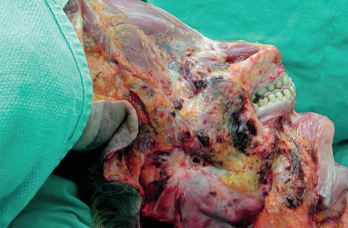
Postmortem hypostatic hemorrhages in a 36-year-old woman who died of multiple drug intoxication and was found in the prone position. Top fig: Left side of neck and face with extensive postmortem hypostasis (lividity). Bottom fig: Hypostatic hemorrhages in the subcutaneous tissues of the left side of the face. Awareness of the pitfalls and artifacts in the neck is essential for a satisfactory and evidence-based approach to interpreting observations of the neck at autopsy.

Fig.1 Top: Left side of neck and face with extensive postmortem hypostasis (lividity). Bottom: Hypostatic hemorrhages in the subcutaneous tissues of the left side of the face.
The postmortem hypostatic hemorrhage is the extravasation of blood into the interstitium when two conditions are met. First, a congested venous plexus is distended with blood due to the gravitational effects of livor mortis. Second, there is loss of vascular integrity of the venous channels due to autolysis or decomposition. When these two factors operate together, the combined effect is the formation of focal perivascular extravasation of blood or extensive interstitial infiltration of blood. The most commonly encountered and trivial examples of this phenomenon are the small, postmortem hemorrhages that occur in the skin in cases of hanging with pronounced lividity in the lower extremities. These postmortem hypostatic petechial hemorrhages (often known as Tardieu spots) are a typical example of postmortem hypostatic hemorrhages.

Latest posts


