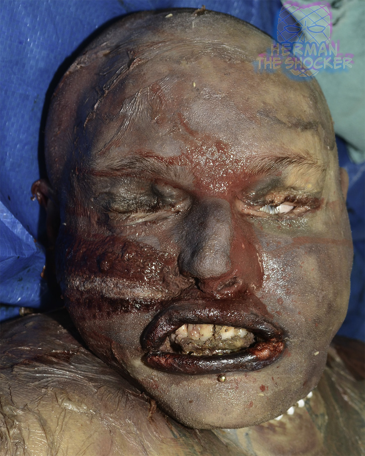
Australia. This middle aged male individual was located at home in an advanced state of decomposition. Postmortem CT showed that the individual had numerous surgical repairs in the form of metal plates and screws on the left frontal-zygomatic suture, left inferior orbit, left maxilla, left mandibular body, mid- and right mandibular symphysis and on the right lateral clavicle. A number of soft tissue piercings were also noted.

Fig.1 Anterior view of the head showing the advanced stage of decomposition.
There are a range of dental implant prosthetics. The pattern of prosthetic repair in the individual presented in this case is indicative of complex reconstructive surgical intervention, which suggests he suffered major head and chest trauma in the past. This repair is likely to be highly individualized and described in detail in medical records, which are likely to include clinical radiographs and/or CT scans. Such evidence may assist in the identification process.

Fig.2 VR images of the skull showing plate fixations. NB: a metal tongue piercing is also visible in the images on the top and bottom right.

Latest posts


