Toronto, Canada. A 34-year-old intoxicated male was found dead in his apartment. On scene inspection, the deceased body was found in the sitting position on a chair, and a massive arterial blood pattern was observed at the scene. First responders and the coroner who visited the scene considered this case as criminally suspicious due to arterial blood patterns on walls and excessive blood pool at the scene.

Fig.1 Deceased in the sitting position on a chair at his residence.
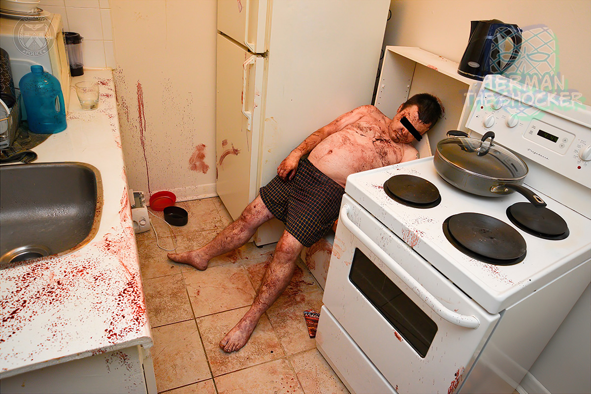
Investigating team that was comprised of police, coroner, a forensic pathologist, and a forensic blood pattern analyst were informed about the presence of an arterial pattern of bleed in the kitchen during the visit at the scene. The presence of an arterial pattern of bleed indicated an injury to an artery. Clothes that were heavily soaked in blood were also found on the floor of the house. Blood smears were also present on the stairs and adjoining walls leading to the deceased’s residence. However, the main medico-legal issues that required were to determine the cause and manner of death.

Fig.2 On inspection of his residence, a large pool of blood was present on the floor of the apartment.
On external examination, a laceration on the left side of the forehead was identified. A detailed examination of the laceration revealed a laceration of the superficial temporal artery (STA) due to blunt impact head trauma. The cause of death was opined as exsanguination due to STA laceration due to blunt impact head trauma. The absence of forced entry into the house and analysis of video of surveillance camera showed that he tripped and hit his head on the edge of a step of the stairs while climbing on it. The scene examination findings, autopsy findings, video of a surveillance camera, and the presence of alcohol in his blood helped in concluding the manner of death as accidental.

Fig.3 Post-mortem CT examination shows no fractures or bleeding in the brain or any other body cavity.
At autopsy, a laceration was present on the left side of the scalp with an underlying transection of the left superficial temporalis artery. A subsequent histological examination of the arterial section established its transection and cellular response to injury. The toxicological analysis report was positive for ethanol, paroxetine, and diazepam. The femoral blood ethanol was 284 mg/100ml. Paroxetine and diazepam concentrations were within therapeutic levels.

Fig.4 Scalp a laceration with associated contusion and superficial temporal artery tear.
After careful perusal of CCTV camera footages, pre-autopsy CT, macroscopic examination of injury, histological examination of the transected artery, and toxicological analysis report, cause of death was given as exsanguination due to laceration of a superficial temporal artery following blunt force head trauma. Ethanol, paroxetine, and diazepam were included as contributing factors.

Fig.5 Sections of the scalp wound showed a laceration involving epidermis and upper dermis and laceration of the superficial temporal artery. (magnification ×20).
Alcohol causes vasodilatation and may enhance the already bleeding vessel. There are few case reports in which the peripheral artery was injured in an alleged attack, and victims were bled to death. In this case, victims were under the influence of alcohol at the time of death. In this case, the deceased was a chronic alcoholic, which resulted in causing fatty change or steatosis in the liver. A fatty change in the liver hampers its capacity to produce the coagulant factors. Thereby non-formation of a blood clot in the injury fails suppression of bleeding, leading to accelerating fatal bleeding.
The manner of death was accidental. Although it is not uncommon to see deaths of alcoholics following scalp trauma, the peculiarity of this case lies in the fact that no evidence of fatal accidental superficial temporal artery has ever been reported in the forensic literature.

Latest posts


