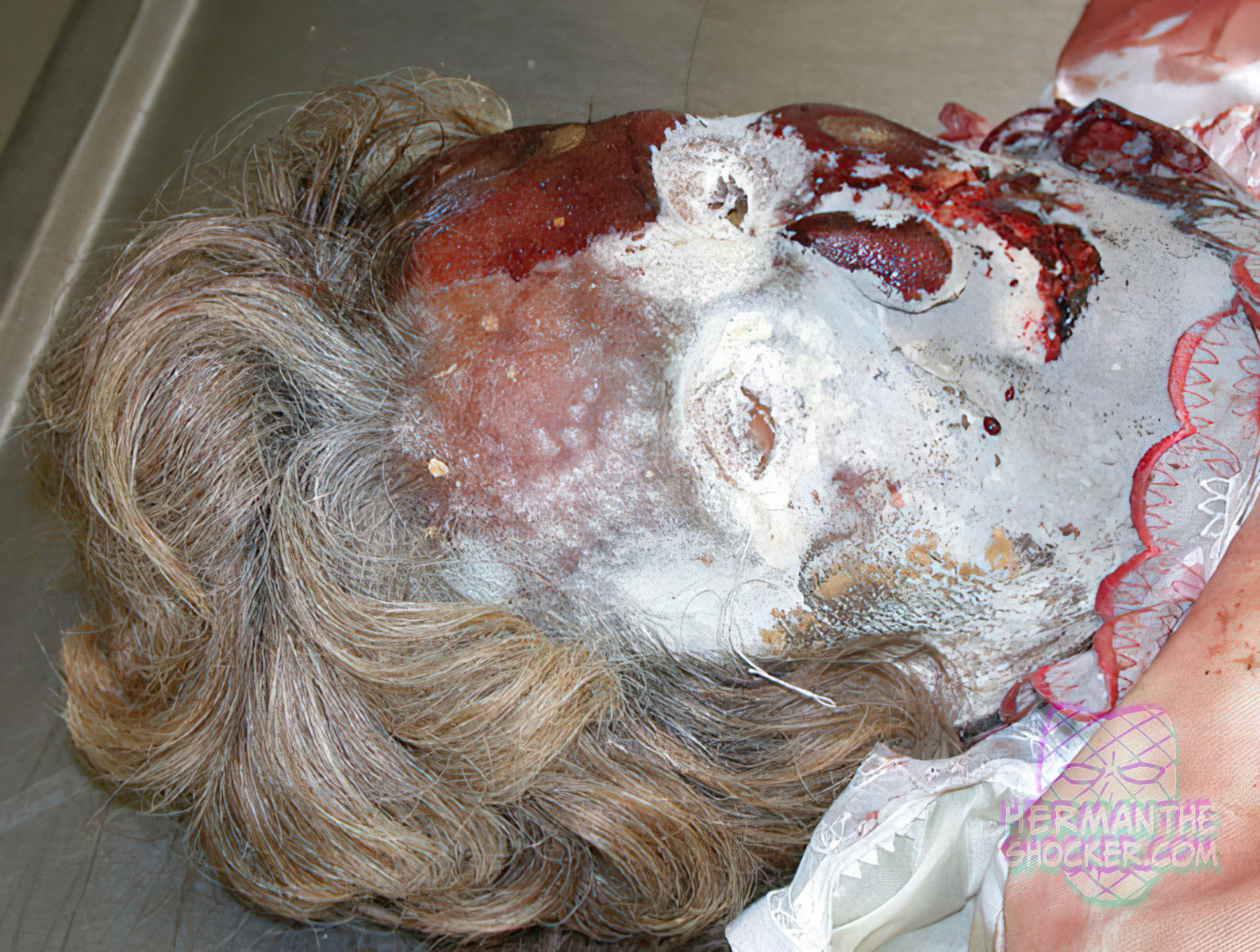
Here we see an extensive whitish-gray fungal colonization on a corpse’s face following a burial time of 7 months. It is often possible to make numerous findings after exhumation despite factors such as autolysis, putrefaction, possible animal predation, fungal colonization, mummification, adipocere, and artifacts (e.g., caused during body recovery). There is no linear correlation between postmortem interval and the detectability of findings, which depends far more on ambient conditions and the diagnostic question being posed. A body may have undergone decomposition to an extent that it is no longer possible to make a targeted assessment.

Fig.1 Extensive whitish-gray fungal colonization following a burial time of 7 months.

Latest posts

This photo shows the effect of a bullet that has grazed the scalp. A bullet graze wound occurs…

This individual died and his body was scavenged by a cat, no further info. The phenomenon of postmortem…

Note the bloating and decompositional blisters as well as the greenish discoloration around the area of the abdomen.…

This individual was strangulated with a rope and then the assailant cut the neck. In a homicide where…
