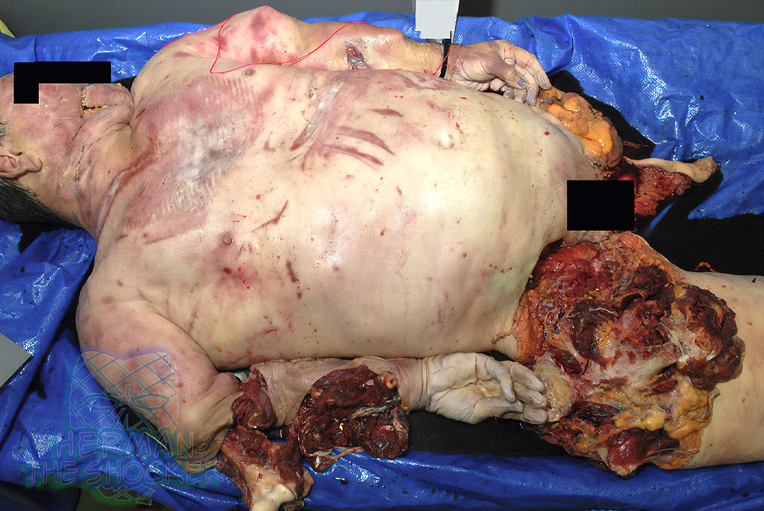
Australia – 2016. Four elderly adults died in a light-plane accident. The light plane crashed into water. Male A: There were multiple abrasions, bruises, and lacerations over the body. There was marked disruption of the head, predominantly around the left to lower aspect of the face. There was a large defect measuring up to 20 cm with multiple skull fragments visible. Diagonal abrasions were seen over the chest, associated with deep bruising. There were multiple bilateral anterior, lateral, and posterior fractures of the ribs. The sternal body was fractured. There was separation between the 3rd and 4th and 8th and 9th thoracic vertebrae associated with disruption of the spinal cord at the level of the cervicomedullary junction. There were multiple fractures of the sacrum and the left and right elements of the pelvis.
Multiple full-thickness defects were seen around the right elbow, back of the forearm, wrist, and hand exposing the disarticulation of the radius at the elbow joint. There was a midshaft compound fracture of the bones of the right forearm. A full-thickness skin defect was seen over the back of the left forearm to wrist, exposing a compound fracture of the radius. The left distal ulna was disarticulated from the wrist mad and protruded through an overlying laceration. There was an extensive laceration to the lateral and anterior aspects of the proximal left thigh. There was a further laceration of the front of the right thigh exposing the distal femur.
There were compound fractures of the left midshaft tibia and fibula associated with an overlying laceration. There was a laceration over the medial aspect of the left ankle, with disarticulation of the medial ankle joint. There was a laceration over the right thigh with exposure of a compound fracture of the distal femur. There was a laceration over the front of the right shin, exposing compound fractures of the tibia and fibula. There was also a laceration over the lateral aspect of the right ankle, exposing the distal fibula, which was fractured. Dissection of the dorsum of each foot showed a small amount of hemorrhage, associated with fractures of the feet.
Male B: Similar to Male A, Male B also had extensive trauma to the body, involving the head, neck, chest, abdomen, pelvis, and limbs. The incomplete left leg (which was fully skeletonized) and right foot were located separately. There was evidence of perimortem trauma to the distal left tibia.
Male C: The only body part of Male C that was recovered (approximately 2 weeks after the aircraft crash) was a partial left pelvis (pubis and ischium were absent), small fragment of sacrum, complete left femur, and complete left tibia. Although the skeletal elements were articulated, there was minimal soft tissue present.
Female D: There were a number of lacerations on the head including those on the right forehead, right lateral eyelid, left upper eyelid, right lateral earlobe (with complete transection of the tissue), left chin to above the right upper lip, right nostril, and right chin. The laceration that extended from the left chin to above the right upper lip was associated with a mandibular fracture and tooth loss. There was extensive disruption and laceration of the anterior groin and genital area through which small bowel loops were extruding. There was also upper and lower limb trauma with degloving injuries and loss of tissue.
There was significant blunt-force trauma to the chest with rib and sternal body fractures, pulmonary lacerations, and prominent alveolar hemorrhage. Within the abdomen there were hepatic, splenic, and mesenteric lacerations. There were cervical and thoracic vertebral body fractures with exposure of the thoracic spinal cord. There was transection of the descending thoracic aorta with both complete and partial tears of the wall.
Although all the deceased were recovered from water, there were considerable differences in both body preservation and the pattern of injuries seen in the four individuals. Interpretation of the circumstances of death based on the analysis of from the remains was complicated by the different times of retrieval of the body parts from the water. The aorta in Male A, Male B, and Female D were transected, consistent with a rapid deceleration injury. Given the severity of the injuries, death of these individuals was likely to have occurred instantaneously. It was not possible from the examination of the injuries to determine where any of the four individuals were positioned in the aircraft.
Postmortem CT images of Male A showed extensive trauma to the anterior and bilateral skull and the body, involving the neck, chest, abdomen, back, pelvis, and limbs. There were fractures of the right proximal humerus, right scapula, pelvis, left neck of femur, left tibia and fibula, right distal femur, right tibia and fibula, and right metatarsals and phalanges. Postmortem CT images of Male B showed fractures of the bones of the face and mandible (the cranial vault was unremarkable), bilateral fractures of the ribs, and fractures of the right radius and ulna, pelvis, and lower limbs.
Postmortem CT images of Female D showed minimal skull disruption compared with the other passengers. The individual had evidence of a Le Fort 2 fracture and fractures of the bilateral mandibular condyle, the right mandibular ramus, and the mandibular symphysis. The cranial vault was unremarkable. Similar to the other passengers, Female D also had extensive post-cranial skeletal trauma.
Latest posts


